
Neuromodulation / Stimulation / Non-ablative Therapies
Neuromodulation is a type of therapy that is used to treat serious, chronic pain and circulatory disorders. The therapy prevents pain impulses from being sent to the brain via the spinal cord. All effects of the therapy are completely reversible. Modulation involves influencing some nerve functions by using electrical impulses (stimulation) or medicines (that enter the central nervous system).
This method has existed since 1972. Since then, it has been much improved thanks to crucial experience and technical developments. Cutting through nerves, which entails permanent damage to the nerves, is something that is now only done in rare, exceptional cases.
This way of relieving pain has only existed since the end of the 1990s. Under local anaesthetic and when the patient is dozing, the flexible radiofrequency catheter is introduced via a small opening near the sacrum, above the anal fold and into the epidural cavity (space between the exterior dura mater of the spinal cord, and the vertebrae), where it is directed towards the nerve root that is causing pain.
Once the catheter has reached its destination, the patient is asked to contribute to the process. Firstly, a weak current is conducted into the painful area, and when the patient feels a tingling sensation in the area of the pain, treatment is carried out using a magnetic field that pulses very quickly (approx. 900,000 times per second) for 300 seconds. These oscillations alter the nerve for a limited period of time, and we therefore describe this process as reversible since it does not cause any lasting damage. The aim of the procedure is to lessen chronic neuropathic pain, which it succeeds at for more than 1.5 years in 70 % of cases.
After the procedure, the catheter is removed again.
As this method is very easy to deal with, it is very popular. It is used in the case of chronic pain originating in the spine, for example, for intervertebral disc protrusions, small prolapses and scarring following operations, but also in the case of spinal stenosis, osteochondrosis, neuroforaminal narrowing, or pains following inflammation and shingles.
Rare unwanted consequences of the intervention may include the following: No guarantee of effectiveness, risk of infection, nerve damage, painful direct muscle stimulation, haematomas and formation of seromas, in addition to intestinal perforation accompanied by peritonitis and injuries to the dura mater, which can cause strong headaches. At our practice we are yet to experience any of these complications, even though we have been carrying out the intervention for several years.
The invasive procedure is carried out in accordance with the guidelines of the International Spine Intervention Society (ISIS).

The great advantage of this minimally invasive procedure is that no open wound is made, no material is left in the spinal canal, and the effects are reversible, meaning that the pain can be selectively targeted. Depending on the indication, we have access to several different catheters for this method.
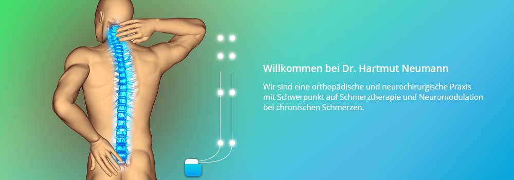
The spinal cord stimulator (SCS) or pain pacemaker is a device that influences the nerves in the spinal cord with an electrode, using weak electric currents. The electrode is introduced into the epidural cavity, through a small opening between the bag that contains the nerves, and the vertebral arch.
The device consists of an electrode and a generator. Using a remote control, the generator can be directed through the skin – the length of impulses, frequency and current strength can be set and adjusted.
Since the 1960s, we have known (thanks to gate control theory) that the spinal cord interprets the impulses that come from the nerves. Some signals are strengthened, while others are almost completely extinguished.
To give you an example: ear clips or the elastic of our trousers should actually cause us constant pain, but they do not. This is because the calls for help from our skin, which are triggered by the constant pressure, are weakened or suppressed by our spinal cord. On the other hand, signals can also be strengthened, which tends to especially happen if we are stressed.
The intervention is carried out in hospital. There are a whole range of approved indications that are recommended by German health insurance companies. These include intractable (which does not respond to conventional therapies) sciatica (leg pain), peripheral arterial occlusive disease (PAOD), and complex regional pain syndromes I and II (CRPS/Sudeck’s atrophy).
Other conditions have also proven to be very good indications for spinal cord stimulation, including chronic pain following a slipped disc operation, stiffening, back pain, post-herpetic neuralgia, stump pain, amputation pain and many others.
Not all types and locations of pain can be treated using SCS, however. Sometimes it can be very difficult to reach the precise location of the pain using the stimulation.
If the patient moves too much too soon after the electrode has been positioned, it can slip and then not have the intended effect. After a few years, the pain pacemaker must be replaced during another small operation, as the battery will run out. If a lot of electricity is used, we therefore place very expensive rechargeable generators under the skin.
Before the generator is implanted, a test stimulation is carried out. Firstly, the electrode is introduced epidurally via a small surgical access hole, under local anaesthetic and light sedation. The patient tests the stimulation for a few days via an external cable. Next, the pacemaker is implanted or, in case the patient is not satisfied, the electrode is removed.
The great advantage of this method is that less medication is needed, and can sometimes even be dispensed with altogether. This can prevent many side effects of the medication (nausea, dizziness, organ damage, constipation etc.). Also, the method is reversible. When the generator is turned off or removed, the symptoms will return. Nerves are not damaged by this.
The risks of the surgery may include the following, among others: No guarantee of effectiveness, risk of infection, nerve damage, painful direct muscle stimulation, formation of haematomas and seromas, electrode slipping leading to loss of stimulation, electrode breakage, failure of the battery or generator, meaning battery must be replaced.
Exciting technological advances mean that there are now plate electrodes that can be implanted under the skin without large surgical interventions, and that can cover a large pain area.
New generators with different new frequency patterns such as high frequencies, volley-like currents and rechargeable batteries are now available, meaning that more and more patients with chronic pain have been helped during the past years. Thanks to advances in technology, the systems have become better and better, and more and more reliable and versatile.
Why not get in touch? We would be more than happy to advise you.
Occipital nerve stimulation (ONS), also known as peripheral nerve stimulation (PNS) of the occipital nerves, is a particular form of nerve stimulation of the nerves at the back of the head, which are situated under the skin. The method has only been established since 2011.
The system consists of one or several probes and a generator. Usually, the small generator is implanted either in the buttocks, the lower abdomen or below the clavicle, in the chest area.
The generator is controlled via a remote control, with which all settings such as current strength, frequency and length of electrical impulses can be changed.
This particular form of stimulation gives hope to patients that suffer from so-called intractable migraines.
Intractable migraines are headaches that cannot be influenced or improved by at least three different medicines for prevention, or for which the side effects of these medicines would be intolerable. Patients continue to suffer from 15 or more migraines (without aura) per month for a period of more than 3 months.
The intervention is associated with minimal risk. Less frequently, the following complications may occur, among others: No guarantee of effectiveness, risk of infection, nerve damage, painful direct muscle stimulation, formation of haematomas and seromas, electrode slipping leading to loss of stimulation, electrode breakage, failure of the battery or generator, meaning battery must be replaced.
If you suffer from this chronic pain, we will be more than happy to advise you about this treatment method.
Ganglion stimulation is a new, very innovative neurostimulation method in pain management therapy. It is used both for patients that experienced pain even after existing procedures, and for new indications.
A four-pole probe is implanted into the ganglions (nerve bundles) that form a thickening of the nerve root inside or outside the spinal canal below the vertebral arches. Here, there is no intermediate layer between the cerebrospinal fluid and the nerves. The probe is therefore able to come very close to the nerve, no matter what position the body is in. The ganglions are the cell bodies of the first nerves that transport the impulses from the site of the pain.
When the spinal ganglion is stimulated, weak electrical impulses are sent to the ganglion of a spinal nerve. These impulses have the effect of reducing the intensity of the pain. Sometimes, a tingling is felt in the former pain site.
They are also the target area of pulsing radiofrequency therapies.
As with classic neurostimulation, the aim is to push the probe through the epidural cavity. The four-pole probe, which is smaller and thinner than an SCS probe, is guided out of this cavity through side openings in the spine towards the ganglions.

The second step is to connect the probe to a generator. Usually, the small generator is implanted either in the buttocks or in the lower abdomen.
The generator is controlled via a remote control, with which all settings such as current strength, frequency and length of electrical impulses can be changed.
The method is used for chronic groin pain, pain following surgery on a groin hernia, for stump and amputation pain, monoradiculopathies (pain that spreads to the arms or legs) but also pain following gynaecological operations, mastectomies (removal of the breasts), chronic regional pain syndrome (CRPS), neuropathic pain following shingles, thoracic operations (on the chest), shoulder and hip operations with nerve injuries, or simple back pain.
Before the generator is implanted, a test stimulation is carried out. Firstly, the electrode is introduced epidurally via a small surgical access hole, under local anaesthetic and light sedation. The patient tests the stimulation for a few days via an external cable. Next, the pacemaker is implanted or, in case the patient is not satisfied, the electrode is removed.
The great advantage of this method is that less medication is needed, and can sometimes even be dispensed with altogether. This can prevent many side effects of the medication (nausea, dizziness, organ damage, constipation etc.). Also, the method is reversible, as when the generator is turned off or removed, the symptoms will return. Nerves are not damaged by this.
The risks of the surgery may include the following, among others: No guarantee of effectiveness, risk of infection, nerve damage, painful direct muscle stimulation, electrode slipping leading to loss of stimulation, formation of haematomas and seromas, electrode breakage, failure of the battery or generator, meaning battery must be replaced.
The method, which has only been approved since 2011, is surgically more challenging than the other methods mentioned. The results of the studies available are promising.
Why not get in touch? We would be more than happy to advise you.
Nowadays, pain pumps are used less and less frequently. A pain pump is a catheter that is linked to a reservoir. The catheter ends up in the central nervous system, directly in the cerebrospinal fluid underneath the meninges (intrathecally), while the reservoir is implanted under the skin above the abdominal muscles. At regular intervals (usually every 4-8 weeks), the reservoir must be filled with different fluids and mixtures via a tube through the skin. The reservoir can be filled with various fluids and mixtures. In the case of intrathecal drug infusion, the medicine, for example, morphine, is injected directly into the cerebrospinal fluid.
The transmission of pain can be inhibited by the use of various medicines on the spinal cord. By introducing the drug infusion into the cerebrospinal fluid in a targeted way, considerably lower doses of medication are needed compared to when tablets or intravenous infusions are administered. This means that the potential side effects for the patient can be minimised too. Patients suffering from chronic pain can benefit from the intrathecal drug infusion, which especially applies to patients that cannot be treated adequately through medication alone (tablets, capsules), or for whom side effects would be intolerable.
Morphine is used in general, but hydromorphine, ziconotides and other substances are also used.
To find out whether the pain is lessened as hoped for, the implantation of the catheter is initially followed by a test phase. During this period, the patient keeps the external medicine pump (outside the body). The dosage of medicine is adjusted according to the patient’s needs. The lessening of the pain is tested under normal everyday conditions.
Nowadays, pumps are more often used to treat muscle spasms. These muscle contractions, which are often painful, occur following a stroke or may even occur early in childhood due to oxygen deprivation during birth.
There are different sizes of reservoir, but most contain 20-50 ml.
The complications specific to this procedure are:
In some cases, the catheter may slip and become blocked; in very rare cases the pump may also stop pumping. Depending on these possible complications, the lessening of the pain can be disrupted or stopped altogether. In these rare cases, a further operation is needed.
In the case of excessively high concentrations, a serious side effect can be the development of apnoea. This means that the patient gradually becomes unconscious and stops breathing. A typical sign that this is happening is an unusually severe contraction of the pupils.
This complication must also be made known to the patient’s family and friends.
Further known side effects to the medicines used are nausea, vomiting and dizziness.
In very rare cases, some people have an allergic reaction to the implant.
It is also a possibility that there will be problems dealing with the programming device or data transmission.
With the implanted system, magnetic resonance tomographies (MRT investigations) may only be carried out following serious consideration and if necessary following consultation.
If a programmable pumping system is being used, a programming device must be available so that a functional check can be carried out at any time.
Why not get in touch? We would be more than happy to advise you.