
Epidural cortisone injections and Epidurography
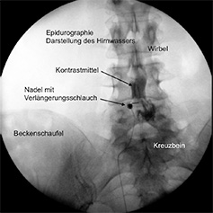
During the treatment of the epidural cavity, a flexible catheter for the use of medicines is placed in the exact location in the spinal canal where it is thought that the damage and therefore the source of the pain are to be found. Under local anaesthetic, the catheter usually is introduced into the spinal canal via an opening in the sacrum. The catheter is made from a particularly easy-to-use plastic pipe with an elastic steel spring at the tip, which means it is easy to steer. During the intervention, the doctor checks the precise location of the catheter using x-ray imaging. Finally, contrast medium is used to show up the epidural cavity (epidurogram) and pain-relieving, anti-inflammatory and anti-swelling drugs are administered.
The treatment is not an open operation, meaning that no general anaesthetic is needed. It is possible to treat several sections of the vertebrae. The indications for targeted segmental administering of medicines into the spinal canal are acute and chronic pain in the case of slipped discs with irritated nerve roots and pain in the area where the nerves are spread out, in addition to narrowing of the spinal canal or of the neuroforamina with stage II PAOD (Claudicatio spinalis), in addition to intractable pain following unsuccessful spine operations, erosive osteochondrosis, and cases of post-herpetic neuralgia that have proven intractable up until now.
If you suffer from the types of pain listed above, we would be more than happy to help you.