
Epiduroscopy
An epiduroscopy or periduroscopy is a percutaneous (through the skin), minimally invasive examination of the epidural cavity (space between the exterior dura mater of the spinal cord, and the vertebrae). By using what are the equivalent of the eyes of a tiny snake, which can make its way around many structures in the spinal canal, we can take a look in almost every corner of the epidural cavity.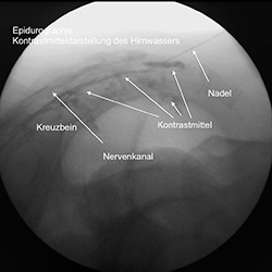
Notwithstanding purposes of the observation of pathology and anatomy, it can also be used to loosen scarring, to position catheters and to administer painkillers directly. Tissue samples can also be taken. The epiduroscopy has both diagnostic and therapeutic uses. The indication range for this innovative method is very broad, ranging from pain syndromes of patients that have already been operated on but are still suffering from their symptoms, to patients that cannot be operated on due to their generally bad state of health, and patients for which conservative (non-surgical) therapy has been unsuccessful until now.
It makes it possible to diagnose and document pains of unclear origin near the spine, especially when it is thought that the cause is in the epidural cavity. It is also used to take samples in the case of inflammatory processes, such as when there is a suspected tumour. Thanks to the epiduroscopy, electrical and mechanical tests can be carried out very close to the spinal cord, catheters can be positioned precisely, and foreign objects can be removed. Above all, scar tissue and adhesions that occur following an operation can be effectively loosened and sometimes even removed. Medicine can be administered very precisely to particular nerves.
The intervention usually lasts around one hour or, in exceptional cases, even less. The endoscope is pushed into the epidural cavity through a small opening in the sacrum, which can be felt under the skin above the anal fold. The patient only feels slight pressure as the endoscope is carefully manoeuvred through the epidural cavity until it reaches the scar tissue or inflammation. X-ray fluoroscopy and the camera images are observed, and the area is cleaned with a powerful water jet so that a good view is possible. At the end, the whole system is removed again and the patient can lie on his or her back once again.
During the first 5-6 hours after the procedure, the following problems can occur: Headaches: if you suffer from headaches, make sure you drink plenty of fluids and take some painkillers, which will usually be effective.
Weak-feeling legs and dizziness: This is a consequence of your blood pressure adjusting, and should you suffer from this, stay in bed.
Pain in the area around the incision: In this case, painkillers should help.